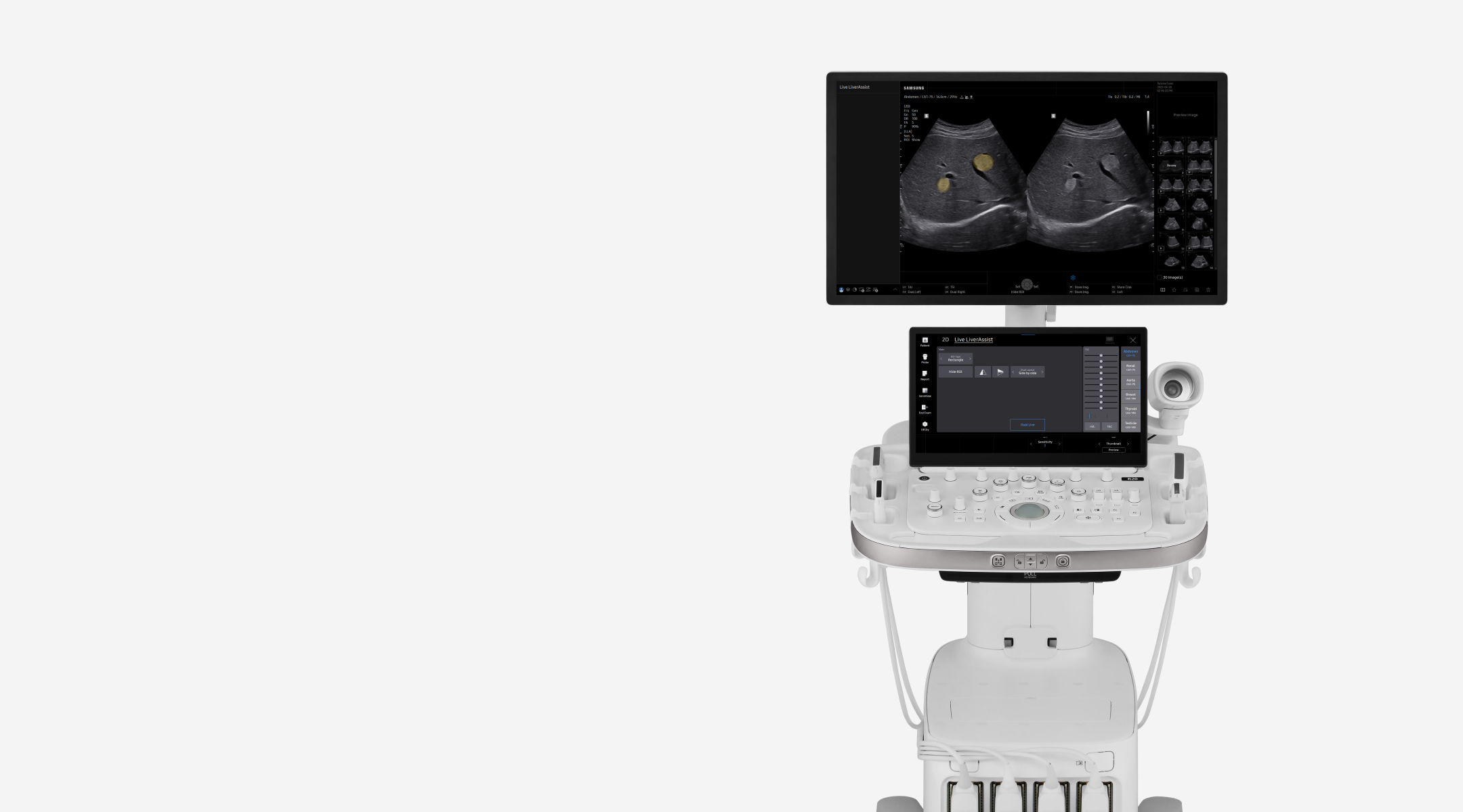
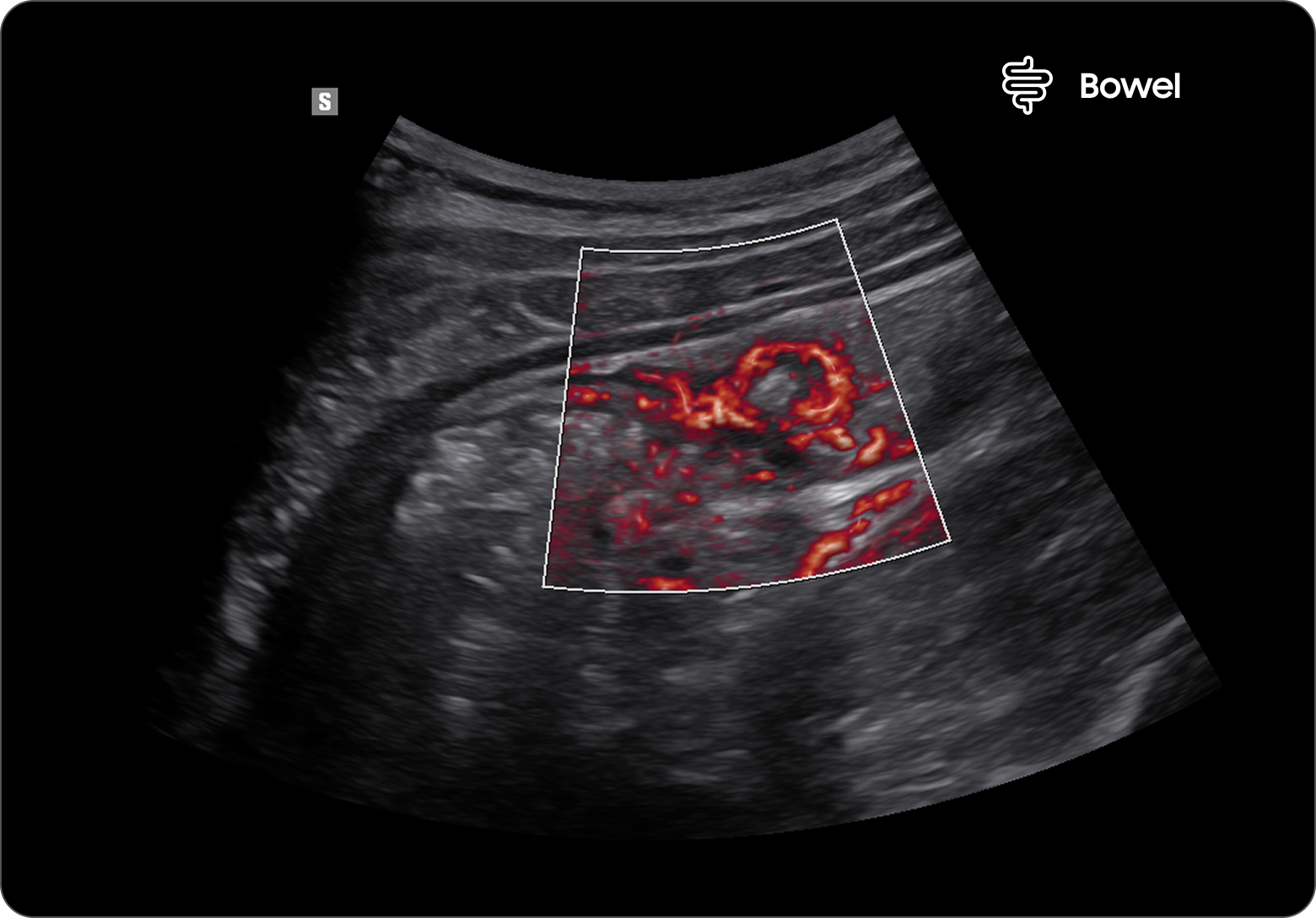
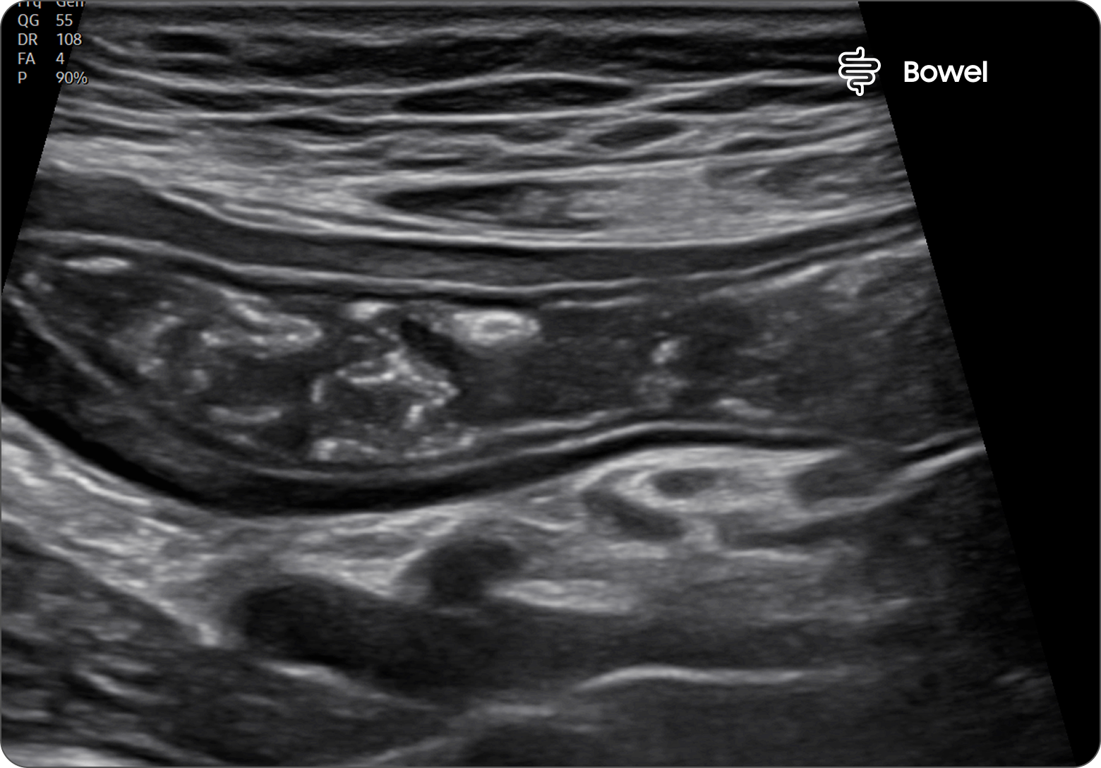
Sorry for the inconvenience.

Chinese users are only allowed to visit websites which complies with the PIPL (Personal Information Protection Law of the People's Republic of China) effective November 1st.

▲ Scan or click the QR code to visit 三星医疗


The personal information of existing Samsunghealthcare.com chinese users will be kept until October 29th and will be safely deleted thereafter.

최적의 환경에서
삼성헬스케어닷컴을 만나보세요

현재 접속하신 브라우저는 지원하지 않습니다.
삼성헬스케어닷컴의 원활한 사용을 위해서는 아래 브라우저 사용을 권장합니다.
브라우저가 설치되어 있지 않은 경우 아이콘을 클릭하여 설치하실 수 있습니다.

현재 사용하시는 브라우저를 확인하려면 아래 사이트를 참고해주세요

https://whatsmybrowser.org/
Ultrasound image of the liver showing hemangioma with MV-Flow highlighting blood flow in red within the lesion

간 내 혈관종 MV-Flow™

Ultrasound image of the gallbladder showing signs of acute cholecystitis with thickened wall and stones

급성담낭염

Ultrasound image of gallbladder and liver hemangioma using 3rd Harmonic for improved clarity and contrast

간 내 혈관종과 담낭 3rd Harmonic Imaging(3차 고조파 이미징)

Ultrasound image of the kidney with MV-Flow showing blue vascular structures within the kidney parenchyma

신장 MV-Flow™

Breast ultrasound image showing an irregular hypoechoic mass with color Doppler flow in orange

형태가 불규칙한 저에코 병변

Breast ultrasound image comparing a mass with and without Live BreastAssist assistance highlighting lesion boundaries

유방 결절 탐지 Live BreastAssist™

Thyroid ultrasound image in trapezoidal view showing the thyroid gland with surrounding neck structures

넓은 시야각을 제공하는 갑상선 영상

Thyroid ultrasound image of a suspicious lesion marked with color Doppler signals in red and blue indicating vascularity

혈류신호를 동반한 악성 의심 병변

Ultrasound image of a finger with DualLive mode showing two comparative views of the soft tissue and bone

손가락 DualLive

Ultrasound image of the brachial plexus with NerveTrack highlighting nerve pathways in yellow

상완신경 NerveTrack™

Ultrasound image of the calf with HQ-Vision showing clear muscle fibers and soft tissue structures

종아리 HQ-Vision™

Ultrasound image of the plantar surface of the foot showing detailed muscle and connective tissue layers

발바닥

Ultrasound image of the liver showing the portal vein and hepatic structures in grayscale

Ultrasound image of the pericallosal artery in a pediatric brain with color Doppler flow in red and blue

뇌량 주변 동맥

Ultrasound image of the bowel with MV-Flow highlighting inflamed areas and blood flow indicating diverticulitis

게실염 MV-Flow™

Ultrasound image of the stomach in trapezoidal view showing layered wall structure and surrounding tissue

넓은 시야각을 제공하는 위 영상

Images(16)

Ultrasound image of the liver showing hemangioma with MV-Flow highlighting blood flow in red within the lesion

간 내 혈관종 MV-Flow™

Ultrasound image of the gallbladder showing signs of acute cholecystitis with thickened wall and stones

급성담낭염

Ultrasound image of gallbladder and liver hemangioma using 3rd Harmonic for improved clarity and contrast

간 내 혈관종과 담낭 3rd Harmonic Imaging(3차 고조파 이미징)

Ultrasound image of the kidney with MV-Flow showing blue vascular structures within the kidney parenchyma

신장 MV-Flow™

Breast ultrasound image showing an irregular hypoechoic mass with color Doppler flow in orange

형태가 불규칙한 저에코 병변

Breast ultrasound image comparing a mass with and without Live BreastAssist assistance highlighting lesion boundaries

유방 결절 탐지 Live BreastAssist™

Thyroid ultrasound image in trapezoidal view showing the thyroid gland with surrounding neck structures

넓은 시야각을 제공하는 갑상선 영상

Thyroid ultrasound image of a suspicious lesion marked with color Doppler signals in red and blue indicating vascularity

혈류신호를 동반한 악성 의심 병변

Ultrasound image of a finger with DualLive mode showing two comparative views of the soft tissue and bone

손가락 DualLive

Ultrasound image of the brachial plexus with NerveTrack highlighting nerve pathways in yellow

상완신경 NerveTrack™

Ultrasound image of the calf with HQ-Vision showing clear muscle fibers and soft tissue structures

종아리 HQ-Vision™

Ultrasound image of the plantar surface of the foot showing detailed muscle and connective tissue layers

발바닥

Ultrasound image of the liver showing the portal vein and hepatic structures in grayscale

Ultrasound image of the pericallosal artery in a pediatric brain with color Doppler flow in red and blue

뇌량 주변 동맥

Ultrasound image of the bowel with MV-Flow highlighting inflamed areas and blood flow indicating diverticulitis

게실염 MV-Flow™

Ultrasound image of the stomach in trapezoidal view showing layered wall structure and surrounding tissue

넓은 시야각을 제공하는 위 영상

자료 및 지원

교육

다양한 초음파 교육 프로그램을 맞춤형으로 제공합니다. 

문의하기

제품 및 솔루션에 대해 궁금하신 점이 있다면 언제든지 문의해 주세요. 

다운로드

세척, 소독, 멸균 방법 안내 및 제품 카탈로그 한눈에 보기 

  • 일부 국가에서는 본 제품 또는 특정 사양이 제공되지 않을 수 있습니다.
  • 제품 판매 및 출하는 규제 기관 승인 후 진행되며, 세부 사항은 지역 영업 담당자에게 문의해 주세요.
  • 본 제품은 의료기기이므로, 사용 전 반드시 사용 설명서를 숙지해 주시기 바랍니다.
  • S-Vue Transducer™는 삼성의 최첨단 단결정 초음파 트랜스듀서명입니다.

    1. 해당 기능은 옵션이며, 추가 비용이 발생 할 수 있습니다.
    2. 음향 진폭은 최대 40%까지 증가하여, 신호 대 잡음비(SNR)가 이에 비례하여 향상될 수 있습니다. 해당 진폭 설정은 인체 안전 기준을 충족하도록 조정되었으며, 3세대 단결정 기술이 CA2-13M 트랜스듀서에 독점 적용되어 영상 품질 향상을 지원합니다.

    Follow Us

    SNS 팔로우하고업데이트 소식을 받아보세요!