Sorry for the inconvenience.

Chinese users are only allowed to visit websites which complies with the PIPL (Personal Information Protection Law of the People's Republic of China) effective November 1st.
▲ Scan or click the QR code to visit 三星医疗


The personal information of existing Samsunghealthcare.com chinese users will be kept until October 29th and will be safely deleted thereafter.

Upgrade browser for full Samsung Healthcare experience

It looks like you may be using a web browser that we don’t support.
Make sure you’re using one of these recommended browsers,
or upgrade your browser by clicking the icons, to get the full Samsung Healthcare experience.

To check which browser you use

https://whatsmybrowser.org/

Select your country or region to find out what content fits your location.

Continue

본문 바로가기

EleV8 to Excellence

Receive exclusive saving offers simply by submitting a demo request for Samsung’s latest ultrasound innovation, V8.

Unifying intelligence and performance

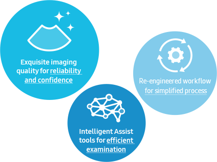
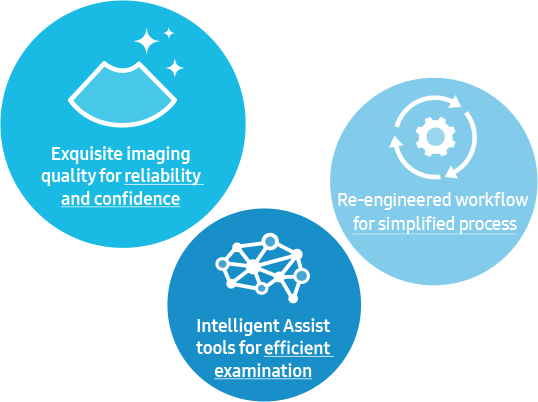
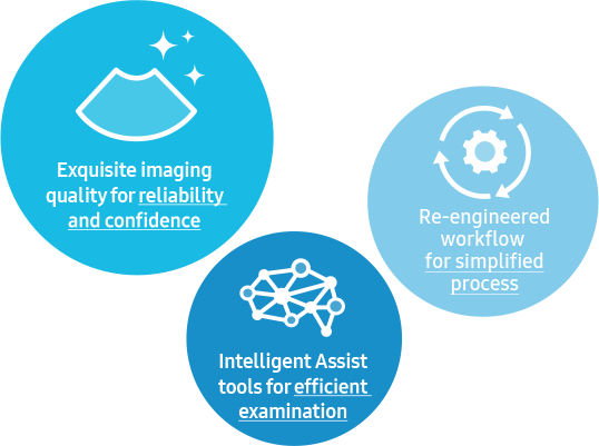
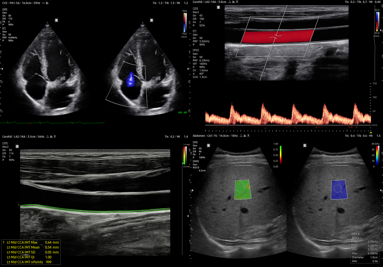
The V8 premium ultrasound system combines exquisite imaging quality powered by Crystal Architecture with efficient, streamlined examination enabled by Intelligent Assist tools, and re-engineered workflow to fulfill the needs of today's busy clinical environment. The sophisticated, ergonomic design showcases Samsung's careful craftsmanship and that comfort-in-use is a high priority for your product experience. We constantly seek new ways to help professionals obtain reliable answers with greater image clarity, enhanced accuracy, and improved work efficiency.

V8 Hands-on

Dr. Julia Aland, a specialist in internal medicine and cardiology, presents the interdisciplinary V8 premium ultrasound system. The focus is on the excellent image and Doppler quality as well as selected functions when used in the fields of internal medicine and cardiology.

Redefined imaging technologies powered by Crystal Architecture

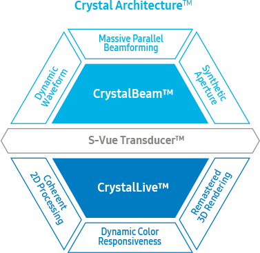
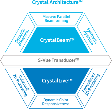
Crystal Architecture, an imaging architecture that combines CrystalBeam and CrystalLive, based upon S-Vue Transducer, provides a crystal clear image. CrystalBeam is a new beamforming technology beneficial in delivering high-quality image resolution and increased uniformity of images. CrystalLive is Samsung’s up-todate ultrasound imaging engine with enhanced 2D image processing, 3D rendering and color signal processing, to offer outstanding image performance and efficient workflow during complex cases.

Exquisite imaging quality for reliability and confidence

Gain insight into the problem based on exceptional image performance powered by Samsung’s core imaging engine, Crystal Architecture. The premium imaging engine combines the benefits of enhanced 2D image processing and detailed expression of color signal processing.

AI
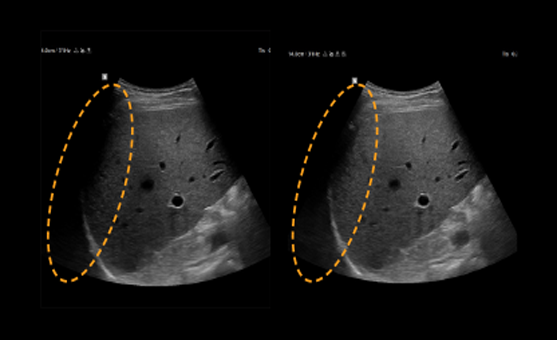
Enhance hidden structures in shadowed regions

Intercostal view / Intercostal view with ShadowHDR

ShadowHDR selectively applies high-frequency and low-frequency of the ultrasound to identify shadow areas such as fetal head or spine where attenuation occurs.

more

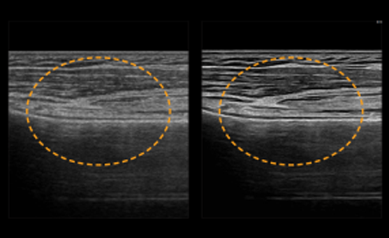
AI
Clean up blurry areas in the image

Quadriceps tendon / Quadriceps tendon with HQ-Vision

HQ-Vison ¹provides clearer images by mitigating the characteristics of ultrasound images that are slightly blurred than the actual vision.

more

AI
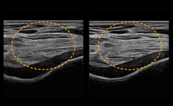
Reduce noise to improve 2D image quality

Biceps tendon / Biceps tendon with ClearVision

ClearVision The noise reduction filter enhances the edge contrast and creates sharp 2D images for optimal diagnostic performance. In addition, ClearVision provides application-specific optimization and advanced temporal resolution in live scan mode.

more

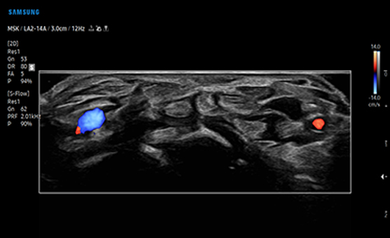
AI
Examine peripheral vessels with directional Power Doppler

Finger flexor tendons with S-Flow

S-Flow, a directional Power Doppler imaging technology, can help to detect even the peripheral blood vessels. It enables accurate diagnosis when the blood flow examination is especially difficult.

more

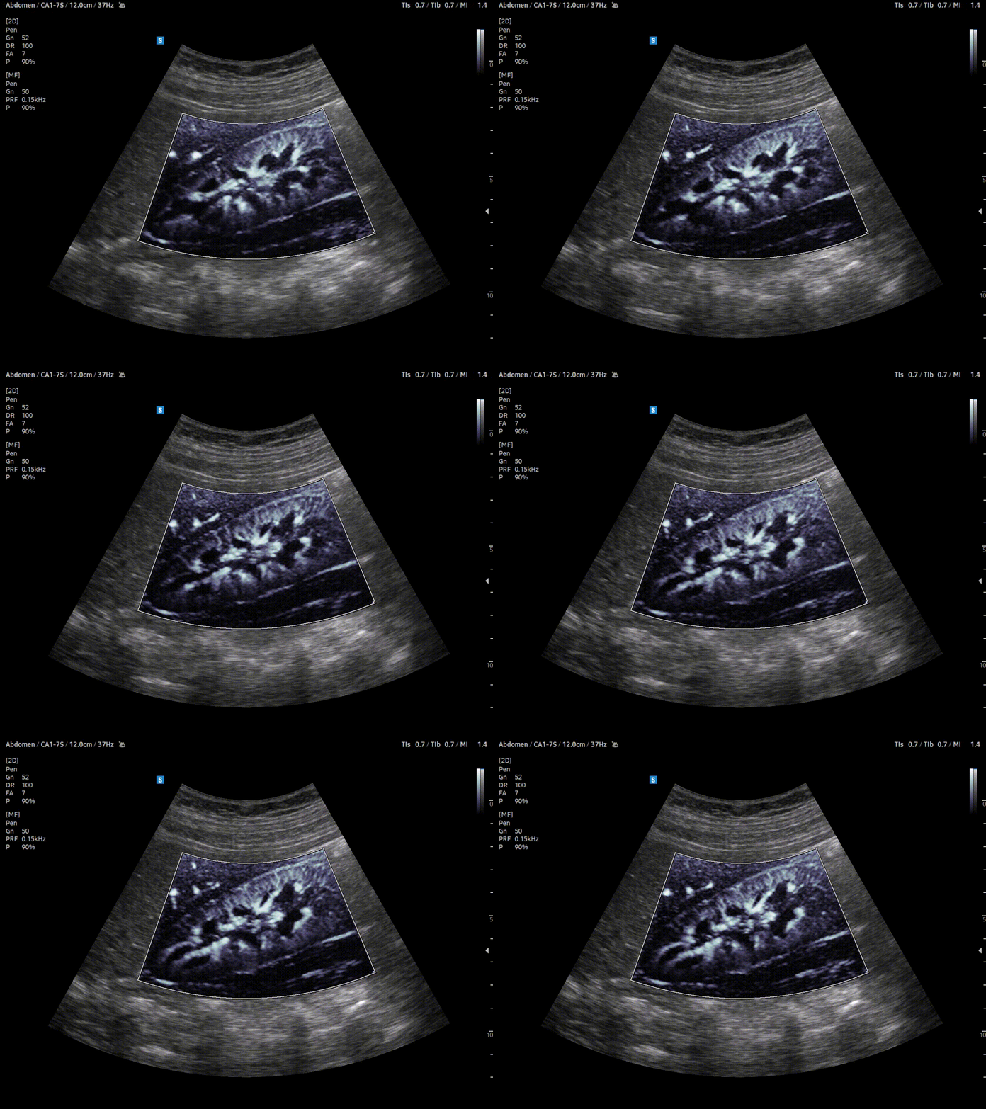
AI
Visualize slow flow in microvascular vessels

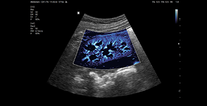
Kidney with MV-Flow

MV-Flow ¹ offers an advanced color imaging for visualizing slow flow of microvascularized structures. High frame rates and advanced filtering enable MV-Flow™ to provide a detailed view of blood flow in relation to surrounding tissue or pathology with enhanced spatial resolution.

more

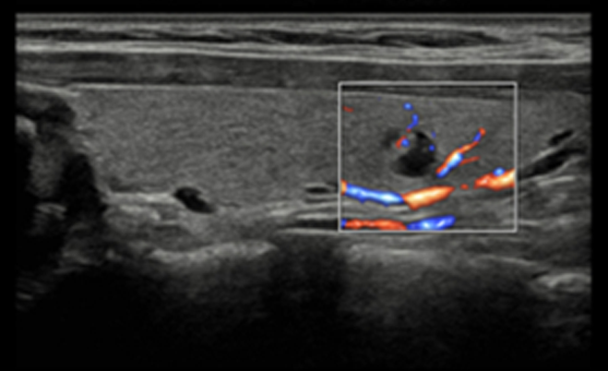
AI
Show blood flow in vessels in a 3D like display

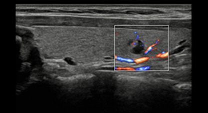
Thyroid nodule (S-Flow with LumiFlow)

LumiFlow ¹ is a function that visualizes blood flow in three dimensional-like to help understand the structure of blood flow and small vessels intuitively.

more

Intelligent Assist tools for efficient examination

Simplify operation and enhance diagnostic confidence with built-in Intelligent Assist features.
V8 supports healthcare professionals with automated features they need to help make decisions.
The system is equipped with a range of tools that help accurately diagnose issues and achieve greater throughput.
For instance, EzHRI™, TAI™, and TSI™ are advanced abdominal dedicated diagnostic features,
that help make accurate clinical assessments by quantifying fatty liver in real time.

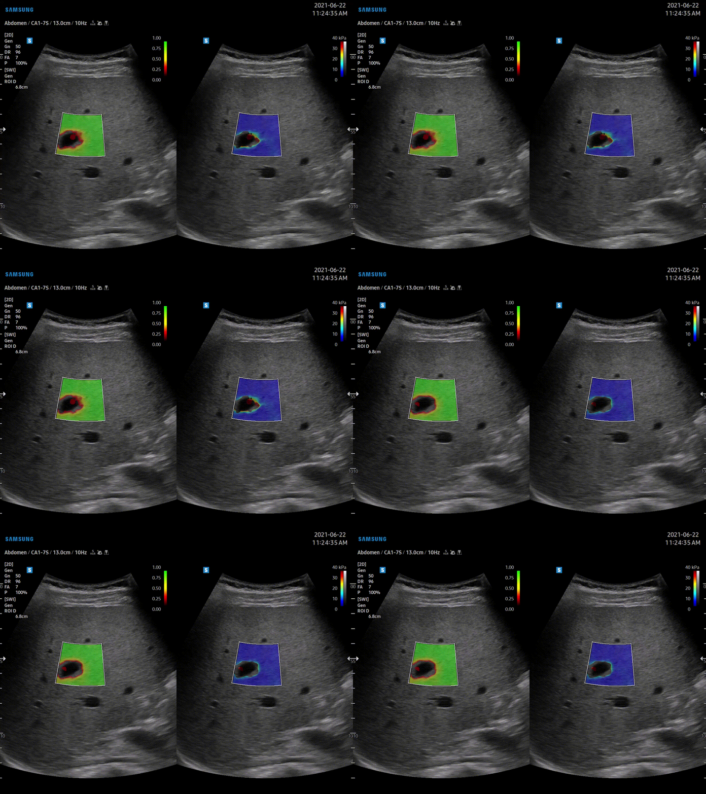
Display and quantify tissue stiffness in a non-invasive method
S-Shearwave Imaging ¹ allows for non-invasive assessment of stiff tissues in various applications. The color-coded elastogram, quantitative measurements, display options, and user-selectable ROI functions are especially useful for accurate diagnosis of breast and liver diseases.
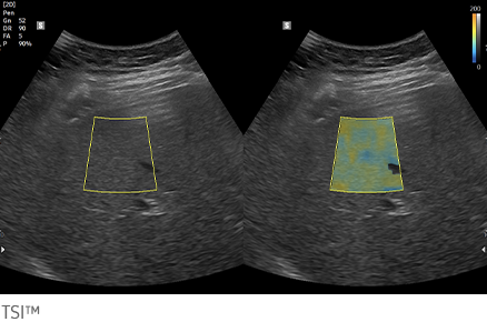
Quantitative measurement of liver fat
with ultrasound signal
TAI™ (Tissue Attenuation Imaging) provides quantitative tissue attenuation measurement to assess steatotic liver changes.
TSI™ (Tissue Scatter distribution Imaging) provides quantitative tissue scatter distribution measurement to assess steatotic liver changes.
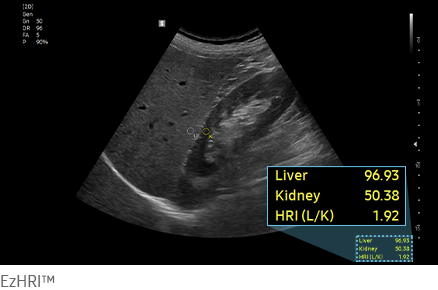
Hepato-renal index with
automated ROI recommendation
HRI (Hepato Renal Index) is an index to quantify steatosis of a liver by comparing echogenicity between liver parenchyma and renal cortex.
EzHRI™ places 2 ROIs on the liver parenchyma and renal cortex and provides HRI ratio.
Perform multi-modality fusion biopsies with high precision
S-Fusion ¹ enables simultaneous localization of a lesion using real-time ultrasound with other volumetric imaging modalities, enabling accurate targeting during interventional and other advanced clinical procedures. Samsung's new Auto Registration helps quickly and precisely fuse the images for increased efficiency.
Contrast Enhanced Ultrasound
CEUS+ ¹ is a contrast enhancement imaging technology that utilizes the characteristics of ultrasound contrast agents. The microbubble contrast agent injected into the body through the vein or alike is subjected to perform nonlinear resonance due to stimulation of ultrasound energy. In addition to the nonlinear signal generated by this method, the ultrasound contrast image is implemented by using the harmonic signal and thus utilized for the diagnosis based on the contrast characteristics over time.
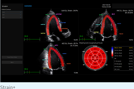
Quantify wall motion of the left ventricle
Strain+ ¹ is a quantitative tool for measuring global and segmental wall motion of the left ventricle (LV). In Strain+, three standard LV views and a Bull's Eye are displayed in a quad screen for easy and quick assessment of the LV function.
Score and report wall motion to determine heart and blood vessel function
StressEcho ¹ package includes wall motion scoring and reporting. It includes exercise StressEcho, pharmacologic StressEcho, diastolic StressEcho and free programmable StressEcho.
Detect functional changes of cardiovascular vessels
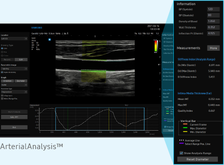
ArterialAnalysis ¹ detects functional changes of vessels, providing measurement values such as the stiffness, intima-media thickness and pulse wave velocity of the common carotid artery. Since the functional changes occur before morphological changes, this technology supports the early detection of cardiovascular disease.
Measure IMT in one click
AutoIMT+ ¹ is a screening tool to analyze a patient's potential risk of cardiovascular disease. It allows easy intima-media thickness measurement of both the anterior and posterior wall of the common carotid by the click of a button.
An automated reporting tool
for heart diagnosis
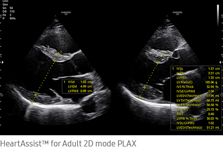
HeartAssist™ ¹, a feature based on Deep Learning technology, provides automatic classification of ultrasound image into measurement views required for heart diagnosis and provides measurement results.
Detect and track nerves automatically with AI technology An automated reporting tool
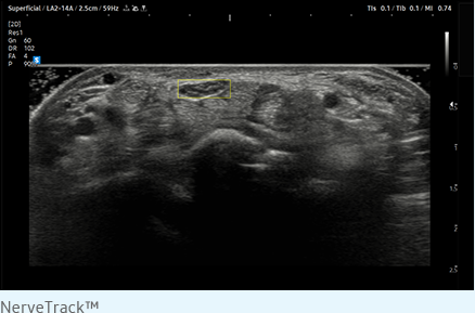
NerveTrack™ ¹, a feature based on Deep Learning technology, detects and provides information of the location of the nerve area in real-time during ultrasound scanning.
Display needle tip clearly
With pinpoint precision, NeedleMate+ ¹ delineates needle location when performing interventions such as nerve blocks. Improved accuracy and efficiency in procedure are possible with beam steering added to NeedleMate+.
AI Analyze selected breast lesions and report breast assessment
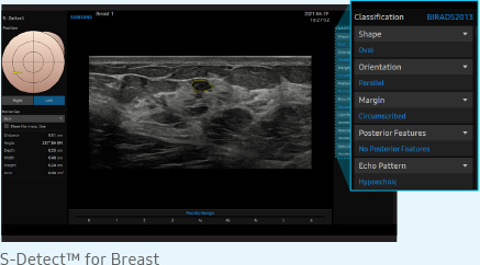
S-Detect for Breast¹, , which analyzes selected lesions in the breast ultrasound study and shows the analysis data, applies BI-RADS ATLAS* (Breast Imaging-Reporting and Data System, Atlas) to provide standardized reporting; and helps diagnosis with the streamlined workflow. * It is a registered trademark of ACR and all rights reserved by ACR.
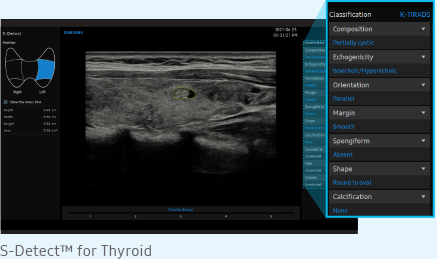
AI Analysis of selected thyroid lesion and reporting for thyroid assessment
S-Detect for Thyroid¹,, which analyzes selected lesions in the thyroid ultrasound study and shows the analysis data, provides standardized reporting based on the ATA, BTA, EU-TIRADS and K-TIRADS* guidelines; and helps diagnosis with the streamlined workflow. * ATA: American Thyroid Association
BTA: British Thyroid Association
EU-TIRADS: European Thyroid Imaging Reporting and Data System
K-TIRADS: Korean Thyroid Imaging Reporting and Data System

Re-engineered workflow and design for a simplified process

Ease your day by streamlining workflow with V8's convenient features and collaborative solutions that reduce multiple tasks into just a few steps and keystrokes. How we display the scan data more easily and precisely is an important focus for the user experience. The ergonomic design makes effective use of the user’s working environment to assure utility and productivity.

Real-time image streaming solution
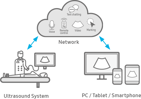
SonoSync is available in PC and smartphone, etc. as a real-time image share solution that allows communication for care guide and training between doctors and sonographers.
In addition, voice chatting, text chatting and real-time marking functions are provided for better communication; and the MultiVue function is included that allows monitoring multiple ultrasound images on a single screen.
* SonoSync is an image sharing solution, not a diagnostic solution.
Build predefined protocols for streamlined process
EzExam+ assign protocols for examinations that are regularly performed in the hospital in order to reduce the number of steps that you have to go through.
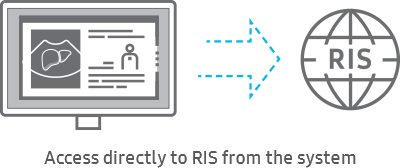
Access to RIS from the browser of the ultrasound system
RIS Browser is a function that improves the workflow in the hospital by allowing access to RIS through the browser embedded in the system for the post process without any need to move to the PC after scanning.
See images in expanded view
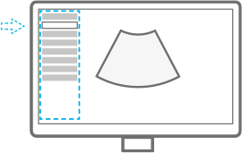
The ultrasound examination can be performed while viewing the images and cines that are expanded at various ratios according to the user preference.
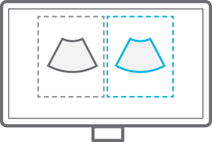
Compare previous and current exam in a side-by-side display
EzCompare automatically matches the image settings, annotations, and bodymarkers from the prior study.
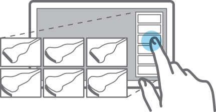
Select transducer and preset combinations in one click
QuickPreset allows the user to select the most common transducer and preset combinations with one touch.
Customize frequently used functions on the touchscreen
TouchEdit , a customizable touchscreen, allows the user to move frequently used functions to the first page.
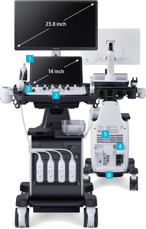
14 inch tilting touch screen
Samsung’s tilting touch screen can be adjusted to accommodate user’s viewing preferences in any scanning environment.
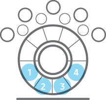
Assign functions to the buttons near the trackball
Depending on the ultrasound inspection items, the functions assigned to the buttons around the trackball can be utilized to reduce the hassle of menu selection.
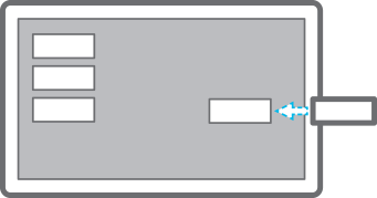
Save image data directly to USB memory
QuickSave function allows image data to be saved directly on USB memory during the exam.
Use the system when AC power is temporarily unavailable
BatteryAssist ¹ provides battery power to the system, enabling users to perform scans when AC power is temporarily unavailable. It also allows to transport the ultrasound system to another location and start to scan right away.
Effective cooling system
An effective airflow system cools down the ultrasound system by constantly letting heat out and reducing fan noise.
Recycled materials
Eco-friendly resin cover is applied to the air vent exterior cover, outlining Samsung's efforts towards a greener tomorrow.

Secure Your Care

Samsung Healthcare Cybersecurity

  • Intrusion Prevention

    Tools for protecting against cyber threats from external attacks

  • Access Control

    Strengthened surveillance for tracking the access of patient information

  • Data Protection

    Encryption functions for safeguarding data whether at-rest or in-transit

LEARN MORE

EleV8 to Excellence

Receive exclusive saving offers simply by submitting a demo request for Samsung’s latest ultrasound innovation, V8.

  • This product, features, options, and transducers are not commercially available in all countries.
  • Sales and Shipments are effective only after the approval by the regulatory affairs.
  • Please contact your local sales representative for further details.
  • This product is a medical device, please read the user manual carefully before use.
  • Optional feature which may require additional purchase.
  • S-Vue Transducer is the name of Samsung’s advanced transducer technology.
  • Strain value for ElastoScan+ is not applicable in Canada and the United States.
  • Recommendations about whether results are benign or malignant in S-Detect are not applicable in the United States.
  • The applicable body parts of NerveTrack™ may vary in the United States and Canada.

Value Package

  • 1 Visualize slow flow in
    microvascularstructures

    Kidney with MV-Flow
  • 2 Show blood flow in vessels
    in a 3D like display

    Thyroid nodule (S-Flow with LumiFlow)
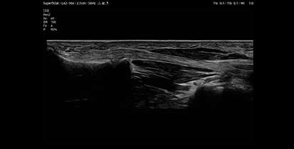
  • 3 Extended Field-of-view
    displaying tool

    MSK with Panoramic
  • 4 DICOM
    (Digital Imaging and Communications in Medicine)

* Depending on the regional regulations and policies, the promotion offers may vary and may not be available in some countries.

The described product information, including features and options, is CE marked. Regulatory approval/clearance status may vary by country. CE 0123